Introduction
Apoptosis, also known as programmed cell death, plays a critical role in maintaining biological homeostasis. It is a tightly regulated process that eliminates unnecessary or damaged cells, thereby facilitating tissue development, immune responses, and the prevention of disease states such as cancer (Elmore, 2007). The ensuing sections delve into the mechanisms, pathways, and significance of apoptosis, with a focus on the latest scientific findings in the field.
Basics of Apoptosis
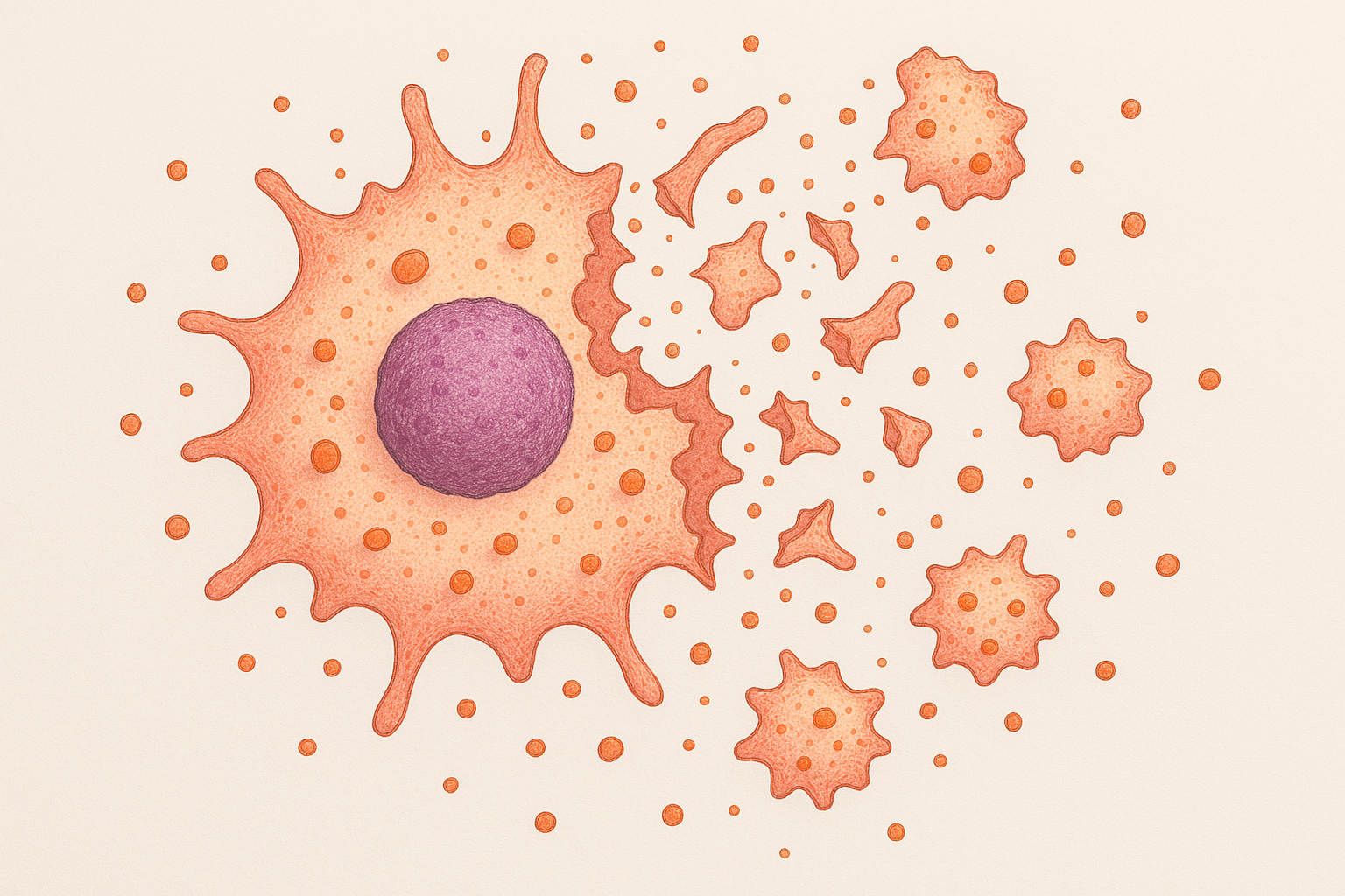
Apoptosis, derived from a Greek term meaning “falling off,” was first described by Kerr, Wyllie, and Currie in 1972 (Kerr, Wyllie & Currie, 1972). Unlike necrosis, which is a form of traumatic cell death resulting from acute cellular injury, apoptosis is a process of cellular self-destruction that is genetically programmed and executed in a highly regulated manner.
Mechanisms and Pathways
The process of apoptosis is mediated by a family of cysteine proteases known as caspases, which once activated, lead to the characteristic morphological and biochemical changes observed during apoptosis (Fuentes-Prior & Salvesen, 2004). The activation of these caspases occurs via two primary pathways – the intrinsic and extrinsic pathways.
The intrinsic pathway, also known as the mitochondrial pathway, is triggered by various intracellular signals such as DNA damage, growth factor deprivation, and oxidative stress. This pathway involves the Bcl-2 protein family, the members of which either promote or inhibit apoptosis (Czabotar et al., 2014).
The extrinsic pathway, also known as the death receptor pathway, is initiated by the binding of death ligands to their corresponding death receptors on the cell surface, leading to the formation of the death-inducing signaling complex (DISC) and subsequent activation of caspases (Ashkenazi & Dixit, 1998).
Significance of Apoptosis
Apoptosis plays an indispensable role in various biological processes. During embryonic development, it helps shape the organism by eliminating unwanted cells. It also plays an important role in the immune system by removing potentially harmful cells, such as self-reactive lymphocytes and virus-infected cells (Fuchs & Steller, 2011).
Moreover, the dysregulation of apoptosis can contribute to various pathological conditions. For instance, excessive apoptosis can lead to degenerative diseases, while insufficient apoptosis can result in the persistence of harmful cells, leading to autoimmune diseases or cancer (Hanahan & Weinberg, 2000).
Recent Advancements and Future Perspectives
Our understanding of apoptosis has significantly expanded over the past few decades. Recent studies have revealed a link between apoptosis and inflammation, providing insights into the role of apoptosis in immune responses and disease pathogenesis (Poon et al., 2014).
Future research in apoptosis is expected to focus on the development of therapeutic strategies that can modulate apoptosis for the treatment of various diseases. For example, pro-apoptotic drugs are being explored for cancer therapy, with the potential to selectively eliminate cancer cells without harming normal cells (Fulda & Debatin, 2006).
Conclusion
In conclusion, apoptosis is a complex and sophisticated process that is crucial for maintaining cellular homeostasis. The study of apoptosis not only enhances our understanding of cell biology but also opens up new avenues for therapeutic interventions. As research in this field continues, it is anticipated that novel strategies for regulating apoptosis will be realized, offering potential breakthroughs in the treatment of various diseases.
References
- Ashkenazi, A., & Dixit, V. M. (1998). Death receptors: signaling and modulation. Science, 281(5381), 1305-1308.
- Czabotar, P. E., Lessene, G., Strasser, A., & Adams, J. M. (2014). Control of apoptosis by the BCL-2 protein family: implications for physiology and therapy. Nature Reviews Molecular Cell Biology, 15(1), 49-63.
- Elmore, S. (2007). Apoptosis: a review of programmed cell death. Toxicologic Pathology, 35(4), 495-516.
- Fulda, S., & Debatin, K. M. (2006). Extrinsic versus intrinsic apoptosis pathways in anticancer chemotherapy. Oncogene, 25(34), 4798-4811.
- Fuentes-Prior, P., & Salvesen, G. S. (2004). The protein structures that shape caspase activity, specificity, activation and inhibition. Biochemical Journal, 384(2), 201-232.
- Fuchs, Y., & Steller, H. (2011). Programmed cell death in animal development and disease. Cell, 147(4), 742-758.
- Hanahan, D., & Weinberg, R. A. (2000). The hallmarks of cancer. Cell, 100(1), 57-70.
- Kerr, J. F., Wyllie, A. H., & Currie, A. R. (1972). Apoptosis: a basic biological phenomenon with wide-ranging implications in tissue kinetics. British Journal of Cancer, 26(4), 239-257.
- Poon, I. K., Lucas, C. D., Rossi, A. G., & Ravichandran, K. S. (2014). Apoptotic cell clearance: basic biology and therapeutic potential. Nature Reviews Immunology, 14(3), 166-180.
Apoptosis Quiz
Test Your Knowledge on Apoptosis!
Answer the following questions to check your understanding of the article on programmed cell death.